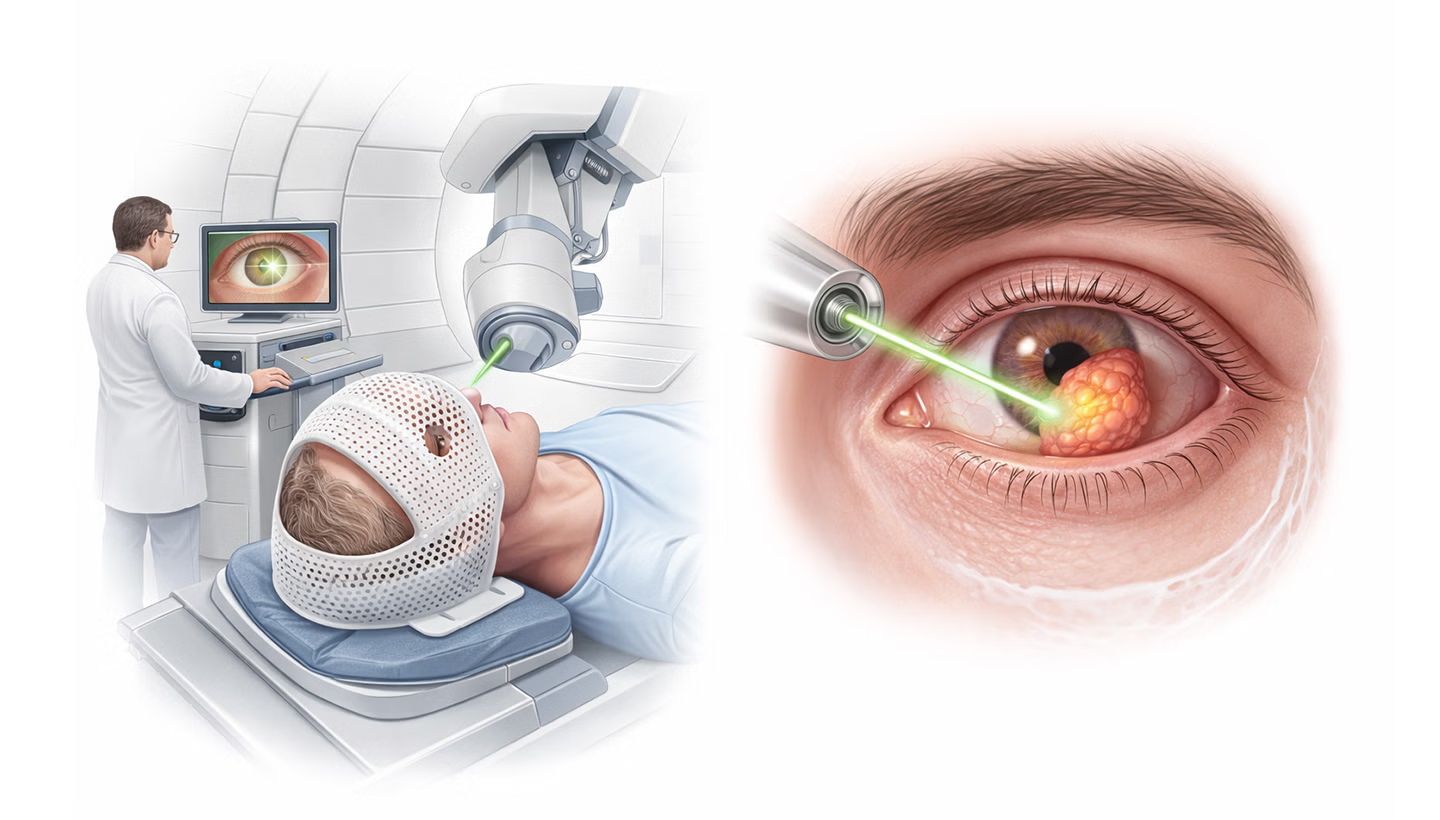
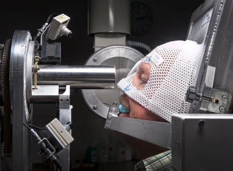
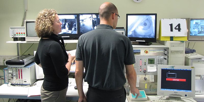
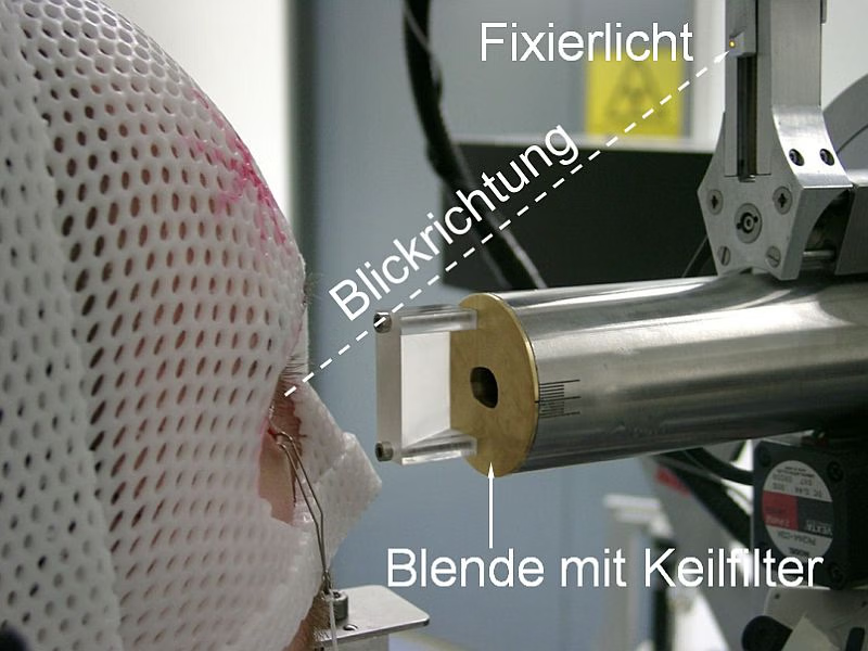
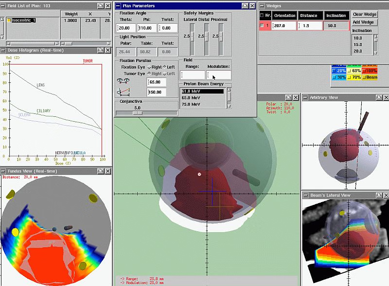
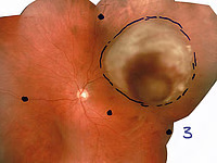
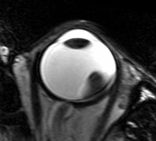
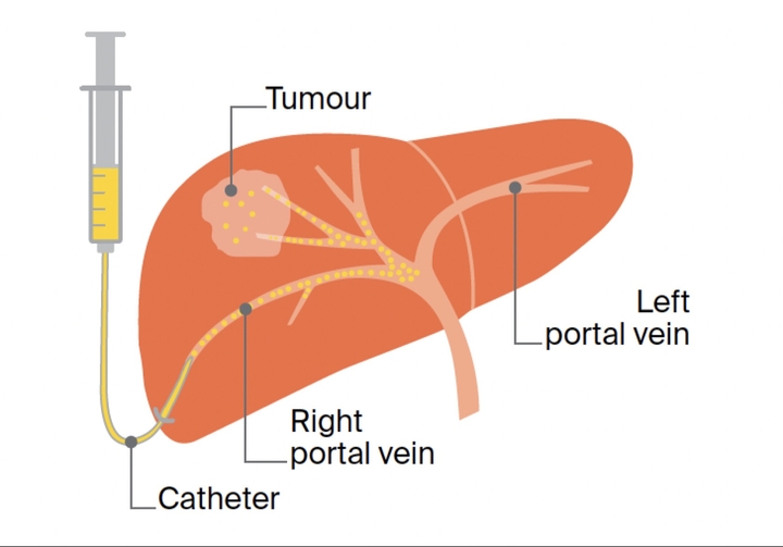
At Charité University Hospital Berlin, we offer proton beam therapy for selected intraocular tumors with the primary goal of destroying the tumor while preserving the eye whenever possible. This treatment is delivered as a joint program between our Department of Radiooncology and Radiotherapy and our Department of Ophthalmology, and it is carried out in cooperation with the Helmholtz-Zentrum Berlin (HZB) at the proton accelerator site in Berlin-Wannsee. The clinic has been operating this ocular proton program since 1998, and with thousands of treated patients, it is are one of the internationally leading centers for proton irradiation of eye tumors. University Hospital Charité Berlin uses ocular proton therapy particularly for uveal melanoma, including choroidal melanoma, and our BerlinProtonen program also covers iris melanomas as well as hemangiomas of the choroid and retina, when an indication for proton irradiation is confirmed. Whether proton therapy is feasible depends strongly on tumor size and exact location in the eye, so not every lesion can be treated with this method. The approach is built around an extremely precise irradiation geometry, allowing doctors to concentrate the dose on the tumor while minimizing dose to adjacent sensitive structures, such as the retina, lens, and optic nerve. In the Charité ocular proton facility, the beam characteristics enable a very steep dose fall-off; for example, it is possible to fully spare the optic nerve even in situations where the tumor is very close to the optic nerve entry, and achieve lid-sparing in more than 95% of treated patients using specially developed lid retractors. The treatment begins with our ocular tumor consultation, which includes a detailed ophthalmologic assessment and documentation, followed by planning diagnostics that include extensive imaging of the ocular fundus. These images are assembled into a fundus composite and are incorporated into the individual proton therapy plan. Because uveal melanomas can metastasize, particularly to the liver, the team also clarifies metastatic status as part of the work-up, with liver assessment performed if it has not already been completed before admission. For the high-precision targeting required in ocular proton therapy, Charité University Hospital Berlin performs a preparatory marker procedure. Doctors suture tantalum position markers onto the sclera to allow exact localization of the tumor at the irradiation position; typically, four markers are placed, and the clip operation takes about 45 minutes and is performed under general anesthesia. After clip placement and precise determination of tumor size and geometry, physicians complete an extensive treatment planning process, which at Charité includes MRI-supported planning for larger eye tumors using specialized software developed in collaboration with the DKFZ Heidelberg and available at our site. The proton irradiation itself is delivered at the cyclotron treatment room. During each treatment session, you are positioned on a precisely controlled treatment chair near the beam exit, with a customized immobilization setup that includes a mask and an individualized bite block. During positioning, X-ray images are used to match the tantalum markers in your eye to the planning data. This setup allows highly reproducible positioning with very high accuracy, and anesthesia is not required for irradiation. For dose shaping, Charité University Hospital Berlin uses a patient-specific aperture matched to the individual tumor size, and the proton beam is directed through this opening to the tumor. The irradiation is painless and very short; the beam delivery itself takes about one minute per fraction, and doctors can interrupt irradiation immediately if the eye leaves the planned position, then resume once correct positioning is restored. In most cases, four irradiation sessions are required within one week, and typically, a plan is made for roughly two weeks in total, including preparation time. With structured follow-up of patients treated at Charité, the hospital reports durable intraocular tumor control in more than 95% of cases and eye preservation in more than 90%, while emphasizing that visual outcomes depend primarily on tumor size and location and that long-term ophthalmologic monitoring remains essential after therapy. After irradiation, the tumor usually regresses slowly over months and remains visible as a scar, and continue regular outpatient controls to verify response and manage any late effects. As with any high-dose radiation treatment in the eye, late complications are possible and are discussed in detail before therapy. These can include radiation-related changes of the retina (radiation retinopathy), optic nerve damage with partial or complete visual loss depending on the received dose, and vascular events such as hemorrhages or perfusion disturbances. The planning focus is to maximize tumor control while minimizing risk to critical ocular structures, but the individual risk profile is determined by the tumor’s proximity to sensitive tissues.
What’s included
Medical service
Examination
clinical history-taking
medical records review
physical examination
consultation with an ophthalmologist
consultation with an anesthesiologist
consultation with a radiation oncologist
Laboratory tests
complete blood count (CBC)
biochemical analysis of blood (kidney and liver function tests, electrolytes)
inflammation blood tests
Diagnosis
eye exam with fundus evaluation
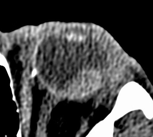
CT of the eye for planning
MRI of the eye (on the indication)
ocular ultrasound
metastasis check
chest X-ray
ECG
Treatment
pre-procedure patient preparation
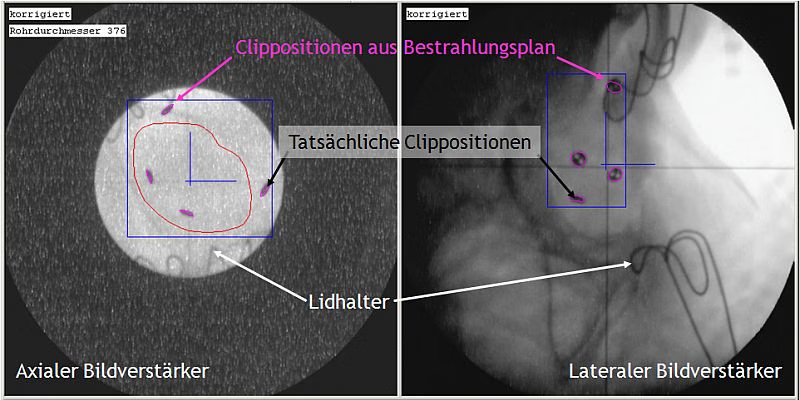
surgical tumor marking (clip operation: tantalum markers are sutured to the sclera (typically 4) under general anesthesia)
3D radiation planning
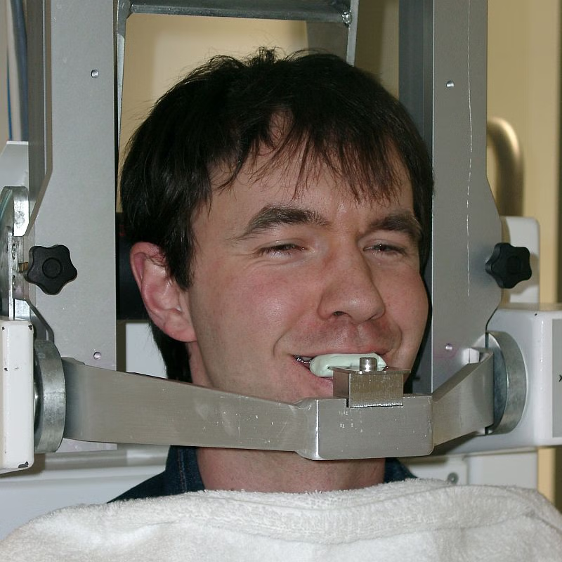
custom immobilization mask fitting
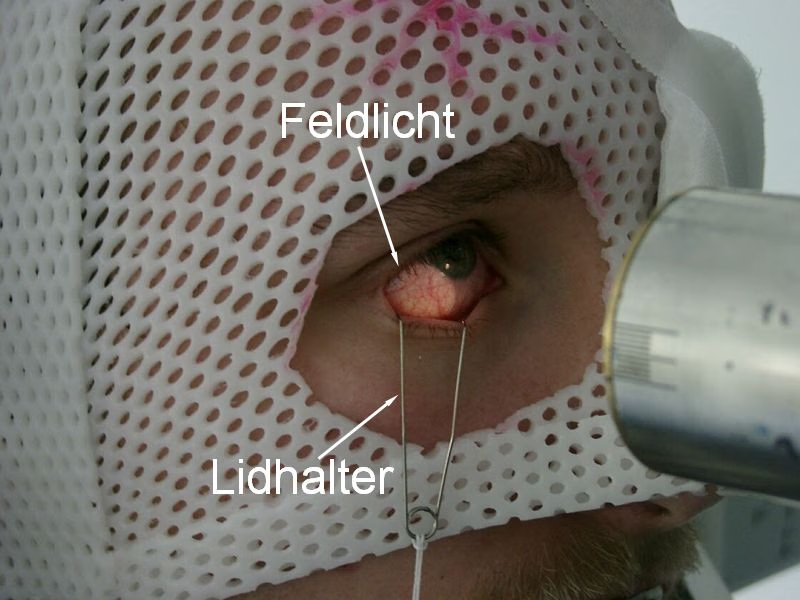
proton beam therapy treatment delivery (eye is locally anesthetized, mask applied, lid holders used, positioning verified with X-ray vs tantalum clips, then irradiation is delivered)
If you're not seeing exactly what you need here, send your custom request. You can discuss the content, specifics, price & timeline to create a personalized plan.
Location
Charitépl. 1, 10117 Berlin, Germany
FAQ
What is the rating of the offer?
Proton Therapy of Eye Tumors at Charité University Hospital Berlin is rated as 9.90 by AiroMedical.