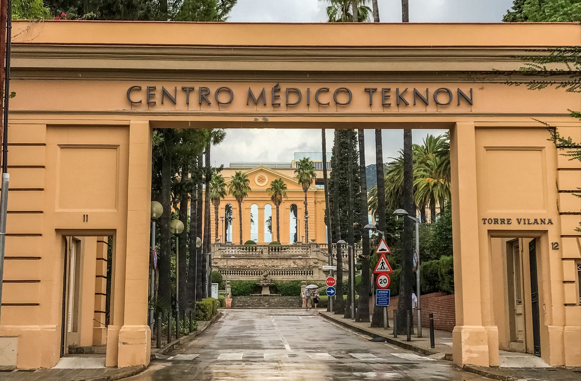
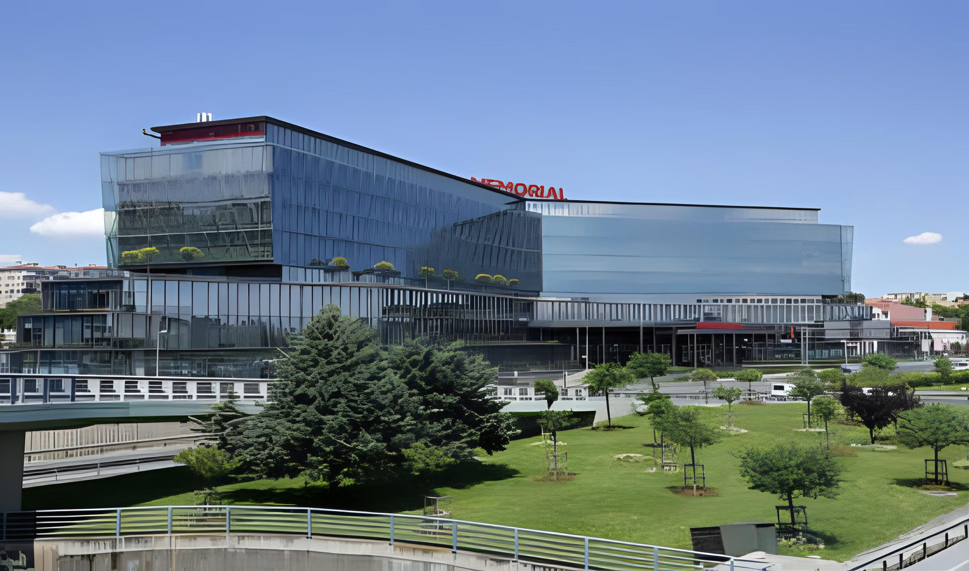
Logo image

Brain tumour treatment

We found 104 clinics & 286 doctors for Brain tumour Worldwide. AiroMedical ranks among 478 hospitals based on qualification, experience, success rate, and awards.

The main characteristics of a benign brain tumour are slower growth and clear separation from normal tissue. They are cured, and the primary treatment method is surgical removal (trepanation, endoscopic intervention or transsphenoidal). Modern techniques include radiosurgery (GammaKnife and CyberKnife) and endovascular embolisation (for vascular neoplasms).

A brain tumour is a group of intracranial neoplasms that can affect cerebral tissue and brain glands (pituitary gland and pineal gland), their membranes, blood vessels or nerves.

The uncontrolled growth of brain structures forms benign brain tumours. They are characterised by slow development and clear boundaries compared to malignant neoplasms. Histological classification of CNS neoplasms defines more than 100 subtypes of primary brain tumours. Doctors determine the kind of the tumour by the cells from which it is formed.

The most common one, which accounts for 50-60% of cases, are glial tumours that develop from glia - auxiliary nervous tissue cells (ependymoma, glioma, astrocytoma).

The next most frequently diagnosed group is benign meningioma (which occurs in 18-28% of cases). It is formed from the hard tissues of meninges. It is followed by a hemangioma arising from brain vascular cells.

Neurofibroma is a benign peripheral nerve tumour that develops from auxiliary cells of nervous tissue (Schwann cells) and cells of the connective tissue (fibroblasts).

Neuro-oncologists distinguish several benign brain tumours: pineocytoma, choroid plexus papilloma, hemangioblastoma, ganglioneuroblastoma, brain angioma and hemangioma.

In addition to non-cancerous neoplasms, the following are formed in the brain:

  • An aneurysm is a bulging and stretching of the aortic walls caused by the thinning of the vessel.
  • Arteriovenous malformations are characterised by abnormal vascular anatomy when venules instead of capillaries follow arterioles.
  • Brain cavernomas consist of pathological cavities separated by sections and filled with blood. They can be located in any part of the brain.
  • A hematoma is an accumulation of blood in tissues due to bleeding. Usually, hematomas result from injuries when the integrity of blood vessels is violated.
Show more

What can cause brain neoplasm?

Scientists have no reliable information that proves the real causes of the disease. Non-cancerous lumps can develop regardless of age. It is also can be congenital. During some studies, oncologists have identified several risk factors that increase the likelihood of developing brain tumours:

  • old age;
  • exposure to radiation (long-term radiation therapy);
  • prolonged contact with harmful toxins;
  • hereditary predisposition;
  • immune system violation;
  • severe brain injuries;
  • living in an area with a dirty environment;
  • undergoing immunosuppressive therapy;
  • some viruses.

Brain tumours are often diagnosed in people with no history of the listed risk factors, so the question of reasons remains open to science.

Nevertheless, the causes of brain hematomas and aneurysms are well known: increased pressure (hypertensive crisis), high intracranial pressure, and brain injuries. At the same time, the causes of cavernomas and arteriovenous malformations are most often abnormalities in the development of brain vessels.

Symptomatic picture of brain tumours

Due to a relatively mild course, these neoplasms can develop asymptomatically for a long time. The main symptom is the occurrence of recurring headaches, which are located in the front and temporal regions of the brain. In addition, the gradual growth of the tumour can cause visual impairment, epileptic seizures and compression of brain tissue.

The general clinical condition worsens with the development of the tumour. It depends on the size, localisation, growth rate and severity of perifocal oedema. According to the statistics, 1-2% of patients aged 45-50 years and older have cerebellar tumours without symptoms and are discovered accidentally during examinations.

Most often, patients complain of the following signs:

  • Headache of a local nature (intense, throbbing), which is challenging to eliminate with the help of painkillers;
  • Constantly increases intracranial pressure;
  • Constant dizziness;
  • Nausea, urge to vomit (regardless of food);
  • Pulsation is possible during palpation;
  • Motor and sensory disorders;
  • Epileptic seizures;
  • Changes in the psyche and personal characteristics (changes in consciousness, memory, attention, etc.);
  • Signs of focal neurology (paresis, impaired vision, hearing, coordination).

The most severe complication is bleeding. In this case, the patient's condition significantly worsens, developing neurological disorders and even death. Intracranial haemorrhage is a severe pathology that can cause damage to the nervous tissue, disruption of the blood supply to the brain and displacement of some brain structures relative to others.

Examination and tests for brain tumours

Diagnosis begins with the initial consultation of a neurosurgeon. He examines your nervous system to check for problems related to the lump. It may include checking:

  • Arm and leg strength;
  • Reflexes such as your knee reflex;
  • Hearing and sight;
  • Skin sensitivity;
  • Balance and coordination;
  • Memory and mental agility using simple questions or arithmetic.

In addition, the physician prescribes standard diagnostic methods, such as blood and urine tests and ECG, to assess the general condition, and accompanied disease.

If a cerebral formation is suspected, the patient will be prescribed the following examinations:

  • Computed tomography of the head (cranial computed tomography, CCT) is a 3D X-ray examination. The body is exposed to a small amount of radiation.
  • The specialist can visualise the blood vessels and thus their abnormalities by administering a contrast agent (CT angiography).
  • Magnetic resonance imaging (MRI) is a gentle method because it works with magnetic fields and radio waves. There is no exposure to radiation. It helps to determine the size and localisation of the brain tumour, as well as to understand how it is connected to the surrounding vessels and structures.
  • Single-photon emission computed tomography (SPECT) and positron emission tomography (PET) allow for studying many processes that underlie the vital activity of the brain, its blood supply and perfusion, the exchange of amino acids, proteins, water, and other features of cerebral metabolism. Currently, SPECT and PET are the main radionuclide methods for diagnosing brain disorders and cerebral tumours.
  • To classify a brain tumour more precisely, the doctor takes a biopsy (tissue sample) and examines it under a microscope. It is done either through the operation or a stereotactic surgical technique. It helps a physician determine the tumour origin and its type.
Show more

Treatment overview for a brain tumour

Not all neoplasms need immediate treatment. For example, if it is tiny and does not cause any symptoms, the doctor uses the wait-and-scan tactic. Here, the development of the tumour is regularly checked using CT or MRI. However, surgery is required if it starts causing discomfort or grows.

The neoplasm is removed as completely as possible. A neurosurgeon will discuss the course of the operation and potential risks with the patient in advance. Surgery is a classic and proven method for brain tumour treatment. If the procedure is performed early, the patient has every chance of a full recovery. A neurosurgeon performs the following interventions:

  • Trepanation of the skull (through access to the skull bone) followed by removal of the tumour. It sometimes is called craniotomy and is an open surgery;
  • Endoscopic intervention through a minimal opening in the head. A neurosurgeon will use a thin probe with the camera to operate inside the skull;
  • Transsphenoidal (through the nasal passages) operation to remove the pineal gland or pituitary gland tumour.

In neurosurgical practice, tumour removal is often performed using laser and ultrasound methods (in cases where the neoplasm is too large or located in an area of ​​the brain inaccessible to the surgeon). Such methods might be MRI-guided laser brain surgery or HIFU device.

Another possible treatment is radiosurgery with GammaKnife and CyberKnife. It is a form of radiotherapy that uses precisely directed ionising radiation to destroy tumour compartments. A high dose affects the formation with high-precision powerful beams with an accuracy of one millimetre at various angles. This way, an effect comparable to surgery is achieved without pain and blood loss.

There is also a special method for brain angiomas, arteriovenous malformations, aneurysms and cavernomas - endovascular embolisation. It is considered a modern, minimally invasive neurosurgical technique. The operation is performed through a single puncture of the artery on the thigh in the groin region. First, the surgeon inserts a thin tubular catheter into the femoral artery, which, under constant X-ray control, is installed in the vessels of the brain directly near the pathological focus. Afterwards, an embolisation substance is introduced through the catheter, which "sticks" the pathological vessels.

However, preserving the brain functions has absolute priority over completely removing the tumour to enable the patient to continue to lead an everyday life. Therefore, neurosurgeons are more likely to accept a residual tumour than permanent damage to a vital brain function. In the case of benign neoplasms, this residual tumour is often observed for years or is treated and stopped by radiosurgery.

If neurological deficits (speech or movement disorders) occur postoperatively, inpatient neurological rehabilitation is helpful.

Suffering from minor limitations or if inpatient rehabilitation is not desired, there are numerous outpatient therapy options to make it easier to get back into everyday life. They include outpatient physiotherapy, occupational therapy, speech therapy, psychosocial care, etc.

In case some brain tumours do not require further treatment, regular follow-up checks and MRI examinations are carried out. The intervals for these checks depend on the type of tumour and the recommendations of your neurosurgeon.

Show more

The prognosis for non-cancerous brain tumours

Experts predict a full recovery if we are speaking about benign, well-defined neoplasm that does not grow into the surrounding tissues. But even benign tumours can recur in 3% of cases.

It should also be said about the 5-year tumour recurrence index depending on where it was localised. The lowest index (3%) is found in those in the skull's vault. Tumours located in the region of the Turkish saddle have an index of 19% and located in the part of the body of the sphenoid bone begin to grow again within five years in 34% of cases. Neoplasms in the area of ​​the wings of the sphenoid bone and sinus have the highest recurrence index. It ranges from 60 to 100%.

In general, all benign neoplasms are treatable with the possibility of complete recovery. However, it is crucial to detect the disease quickly, not ignore the symptoms, and consult a specialist. A responsible attitude to your health and timely diagnosis will help to cope with a tumour.

FAQ

Is brain tumour cancer?

Some brain tumours are non-cancerous (benign), and some cancerous (malignant). Benign neoplasms tend to stay in one place and do not spread. Therefore, it will not usually come back if a neurosurgeon can safely remove all tumours during surgery. Non-malignant brain formations include vascular neoplasms (angioma, arteriovenous malformations, aneurysms) and hematomas

Can the prognosis for brain tumour treatment be different?

Of course, each case is individual, and many factors influence the prognosis. First, many types of tumours have different characteristics (size, localisation, origin). The general condition and age of the patient also play a significant role. And, of course, the prognosis is affected by timely diagnosis and a correctly selected treatment regimen.

Is surgery the best option for brain tumours?

Yes, surgery is the method of choice for brain tumours. Surgical removal allows you to eliminate the problem, often without repeated relapses and complications. However, there are cases when surgery is contraindicated, so neurosurgeons use other methods.

Can I treat a brain tumour without surgery?

Nowadays, there are a significant number of modern techniques that treat brain tumours without the need for surgical intervention. But, first, it is essential in case of contraindications or difficult access to neoplasm. The method of choice is radiosurgery with CyberKnife and GammaKnife.

Is brain tumour treatment abroad more effective?

Many patients with brain neoplasms prefer treatment in leading clinics abroad. Brain tumour diagnosis and treatment methods abroad are characterised by a comprehensive approach and the use of high-tech equipment. And neurosurgeons have much more internship and practice experience in American and European medical institutions.

Where can I get Brain tumour treatment?

Germany, Poland, Turkey, Spain, Lithuania are among the best for Brain tumour treatment.